 akash.verma@cenmed.com
 (732) 447-1115

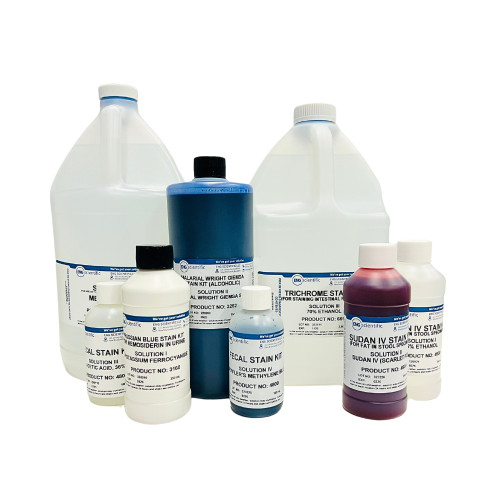
A stain used in histology for the differentiation of melanins and lipofuscins.
CONTENTS: Nile Blue A, Sulfuric Acid
This solution is made from certified dyes.
FOR IN VITRO DIAGNOSTIC USE ONLY.
RECOMMENDED PROCEDURE: Deparaffinize and hydrate to distilled water. Stain 20 minutes. Wash 10 - 20 minutes in running water. Mount in glycerol gelatin.
RESULTS: Lipofuscins - dark blue or green - blue; melanins - dark green. Cytoplasms: muscle - pale green, red corpuscles - greenish yellow to greenish blue, myelin - green to deep blue, nuclei - not stained.
- UPC:
- 12171505
- Condition:
- New
- HazmatClass:
- No
- WeightUOM:
- LB
- MPN:
- 9440